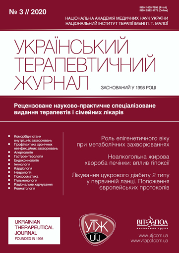
Фактори кардіоваскулярного ризику у хворих на неалкогольну жирову хворобу печінки та їхній зв’язок з мікробіотою кишечника
DOI:
https://doi.org/10.30978/UTJ2020-3-74Ключові слова:
неалкогольна жирова хвороба печінки, чинники кардіоваскулярного ризику, кишкова мікробіота.Анотація
Неалкогольна жирова хвороба печінки (НАЖХП) — одна з основних причин хронічних захворювань печінки, зростання частоти яких спостерігається у світі та зокрема в Україні. Останні епідеміологічні моделі прогнозують прогресивне зростання загальної кількості випадків НАЖХП та її нозологічних одиниць: неалкогольного стеатозу і стеатогепатиту. Актуальність проблеми НАЖХП зумовлена не лише несприятливими епідеміологічними тенденціями, а й соціально-економічними наслідками: підвищенням ризику загальної смертності та смертності, спричиненої серцево-судинними захворюваннями. Встановлено, що НАЖХП асоційована зі збільшенням поширеності традиційних чинників ризику серцево-судинних захворювань, зокрема цукрового діабету 2 типу та ожиріння, але жирова дистрофія печінки є предиктором кардіоваскулярних подій незалежно від її взаємозв’язку з традиційними чинниками ризику.Механізми, за допомогою яких НАЖХП підвищує ризик розвитку серцево-судинних захворювань, комплексні із залученням різноманітних багаторівневих функціональних і структурних шляхів. Однак патогенетичні ланки атерогенезу, асоційовані з розвитком жирової дистрофії печінки, остаточно не встановлено. З огляду на ланки патогенезу НАЖХП, до них, імовірно, належать генетичні чинники, дисфункція жирової тканини, порушення мікроциркуляції та ендотеліальна дисфункція, а також мікробіологічні особливості. Останні відіграють важливу роль у патофізіологічних процесах, які призводять до розвитку серцево-судинної патології у хворих на НАЖХП та які, можливо, мають спільні ланки із патофізіологічними змінами, котрі спостерігаються при формуванні жирової дистрофії печінки. Перспективними напрямами досліджень є з’ясування патофізіологічної ролі кишкової мікробіоти в розвитку як жирової дистрофії печінки, так і чинників кардіоваскулярного ризику, уточнення комплексної взаємодії мікробіоти, метаболічних та імунних каскадів, а також вивчення можливості їх корекції за допомогою як медикаментозних (пробіотики та інші лікарські засоби), так і немедикаментозних заходів.
Посилання
Al-Obaide MAI, Singh R, Datta P. et al. Gut Microbiota-Dependent Trimethylamine-N-oxide and Serum Biomarkers in Patients with T2DM and Advanced CKD // J. Clin. Med. — 2017. — 6. doi: 10.3390/jcm6090086.
Alkhouri N, Tamimi TA, Yerian L. et al. The inflamed liver and atherosclerosis: a link between histologic severity of nonalcoholic fatty liver disease and increased cardiovascular risk // Dig. Dis. Sci. — 2010. — 55. — P. 2644 — 2650. doi: 10.1007/s10620-009-1075-y.
Arpaia N, Campbell C, Fan X. et al. Metabolites produced by commensal bacteria promote peripheral regulatory T-cell generation // Nature. — 2013. — 504. — P. 451 — 455. doi: 10.1038/nature12726.
Atarashi K, Tanoue T, Oshima K. et al. Treg induction by a rationally selected mixture of Clostridia strains from the human microbiota // Nature. — 2013. — 500. — P. 232 — 236. doi: 10.1038/nature12331.
Ballestri S, Lonardo A, Bonapace S. et al. Risk of cardiovascular, cardiac and arrhythmic complications in patients with non-alcoholic fatty liver disease // World J. Gastroenterol. — 2014. — 20. — P. 1724 — 1745. doi: 10.3748/wjg.v20.i7.1724.
Bilotta AJ, Ma C, Huang X. et al. Microbiota metabolites SCFA promote intestinal epithelial repair and wound healing through promoting epithelial cell production of milk fat globule-EGF factor 8 // The Journal of Immunology. — 2018. — 200. — P. 53.17 — 53.17.
Brandsma E, Kloosterhuis NJ, Koster M. et al. A Proinflammatory Gut Microbiota Increases Systemic Inflammation and Accelerates Atherosclerosis // Circ Res. — 2019. — 124. — P. 94 — 100. doi: 10.1161/circresaha.118.313234.
Broeders EP, Nascimento EB, Havekes B. et al. The Bile Acid Chenodeoxycholic Acid Increases Human Brown Adipose Tissue Activity // Cell. Metab. — 2015. — 22. — P. 418 — 426. doi: 10.1016/j.cmet.2015.07.002.
Brouwers M, Simons N, Stehouwer CDA. et al. Relationship Between Nonalcoholic Fatty Liver Disease Susceptibility Genes and Coronary Artery Disease // Hepatol. Commun. — 2019. — 3. — P. 587 — 596. doi: 10.1002/hep4.1319.
Byrne CD, Targher G. Ectopic fat, insulin resistance, and nonalcoholic fatty liver disease: implications for cardiovascular disease // Arterioscler Thromb Vasc Biol. — 2014. — 34. — P. 1155 — 1161. doi: 10.1161/atvbaha.114.303034.
Calori G, Lattuada G, Ragogna F. et al. Fatty liver index and mortality: the Cremona study in the 15th year of follow-up // Hepatology. — 2011. — 54. — P. 145 — 152. doi: 10.1002/hep.24356.
Cetindagli I, Kara M, Tanoglu A. et al. Evaluation of endothelial dysfunction in patients with nonalcoholic fatty liver disease: Association of selenoprotein P with carotid intima-media thickness and endothelium-dependent vasodilation // Clin. Res. Hepatol. Gastroenterol. — 2017. — 41. — P. 516 — 524. doi: 10.1016/j.clinre.2017.01.005.
Chandrasekharan K, Alazawi W. Genetics of Non-Alcoholic Fatty Liver and Cardiovascular Disease: Implications for Therapy? // Front Pharmacol. — 2019. — 10. — P. 1413. doi: 10.3389/fphar.2019.01413.
Chmielewski P. Leukocyte count, systemic inflammation, and health status in older adults: A narrative review // Anthropological Review. — 2018. — 81. — P. 81 — 101. doi: https://doi.org/10.2478/anre-2018-0007.
De Preter V, Coopmans T, Rutgeerts P. et al. Influence of long-term administration of lactulose and Saccharomyces boulardii on the colonic generation of phenolic compounds in healthy human subjects // J. Am. Coll. Nutr. — 2006. — 25. — P. 541 — 549. doi: 10.1080/07315724.2006.10719570.
Deloukas P, Kanoni S, Willenborg C. et al. Large-scale association analysis identifies new risk loci for coronary artery disease // Nat. Genet. — 2013. — 45. — P. 25 — 33. doi: 10.1038/ng.2480..
Dongiovanni P, Petta S, Maglio C. et al. Transmembrane 6 superfamily member 2 gene variant disentangles nonalcoholic steatohepatitis from cardiovascular disease // Hepatology. — 2015. — 61. — P. 506 — 514. doi: 10.1002/hep.27490.
EASL. EASL policy statement on food, obesity and non-alcoholic fatty liver disease (NAFLD) In: EASL, ed, 2019.
Ekstedt M, Hagstrom H, Nasr P. et al. Fibrosis stage is the strongest predictor for disease-specific mortality in NAFLD after up to 33 years of follow-up // Hepatology. — 2015. — 61. — P. 1547 — 1554. doi: 10.1002/hep.27368.
Erdmann J, Kessler T, Munoz Venegas L. et al. A decade of genome-wide association studies for coronary artery disease: the challenges ahead // Cardiovasc. Res. — 2018. — 114. — P. 1241 — 1257. doi: 10.1093/cvr/cvy084.
Eslam M, Valenti L, Romeo S. Genetics and epigenetics of NAFLD and NASH: Clinical impact // J. Hepatol. — 2018. — 68. — P. 268 — 279. doi: 10.1016/j.jhep.2017.09.003.
Estes C, Razavi H, Loomba R. et al. Modeling the epidemic of nonalcoholic fatty liver disease demonstrates an exponential increase in burden of disease // Hepatology. — 2018. — 67. — P. 123 — 133. doi: 10.1002/hep.29466.
Ferrell JM, Boehme S, Li F. et al. Cholesterol 7alpha-hydroxylase-deficient mice are protected from high-fat/high-cholesterol diet-induced metabolic disorders // J. Lipid Res. — 2016. — 57. — P. 1144 — 1154. doi: 10.1194/jlr.M064709.
Francque SM, van der Graaff D, Kwanten WJ. Non-alcoholic fatty liver disease and cardiovascular risk: Pathophysiological mechanisms and implications // J. Hepatol. — 2016. — 65. — P. 425 — 443. doi: 10.1016/j.jhep.2016.04.005.
Ghazalpour A, Cespedes I, Bennett BJ. et al. Expanding role of gut microbiota in lipid metabolism // Curr. Opin. Lipidol. — 2016. — 27. — P. 141 — 147. doi: 10.1097/mol.0000000000000278.
Guss JD, Horsfield MW, Fontenele FF. et al. Alterations to the Gut Microbiome Impair Bone Strength and Tissue Material Properties // J. Bone Miner Res. — 2017. — 32. — P. 1343 — 1353. doi: 10.1002/jbmr.3114.
Haring R, Wallaschofski H, Nauck M. et al. Ultrasonographic hepatic steatosis increases prediction of mortality risk from elevated serum gamma-glutamyl transpeptidase levels // Hepatology. — 2009. — 50. — P. P. 1403 — 1141. doi: 10.1002/hep.23135.
Ismaiel A, Dumitraşcu DL. Cardiovascular Risk in Fatty Liver Disease: The Liver-Heart Axis-Literature Review // Front Med. (Lausanne). — 2019. — 6. — P. 202. doi: 10.3389/fmed.2019.00202.
Jepsen P, Vilstrup H, Mellemkjaer L. et al. Prognosis of patients with a diagnosis of fatty liver-a registry-based cohort study // Hepatogastroenterology. — 2003. — 50. — P. 2101 — 2104.
Kazemian N, Mahmoudi M, Halperin F. et al. Gut microbiota and cardiovascular disease: opportunities and challenges // Microbiome. — 2020. — 8. — P. 36. doi: 10.1186/s40168-020-00821-0.
Kitai T, Tang WHW. Gut microbiota in cardiovascular disease and heart failure // Clin. Sci. (Lond). — 2018. — 132. — P. 85 — 91. doi: 10.1042/cs20171090.
Kurinna О. Pidsumky piatoi shchorichnoi zustrichi Paris NASH meeting (Ukr) // Suchasna hastroenterolohiia [Modern gastroenterology]. — 2019. — 5 (19). — P. 108 — 112. doi: https://doi.org/10.30978/MG-2019-5-108.
Lauridsen BK, Stender S, Kristensen TS. et al. Liver fat content, non-alcoholic fatty liver disease, and ischaemic heart disease: Mendelian randomization and meta-analysis of 279 013 individuals // Eur. Heart J. — 2018. — 39. — P. 385 — 393. doi: 10.1093/eurheartj/ehx662.
Lazar V, Ditu LM, Pircalabioru GG. et al. Aspects of Gut Microbiota and Immune System Interactions in Infectious Diseases, Immunopathology, and Cancer // Front Immunol. — 2018. — 9. — P. 1830 doi: 10.3389/fimmu.2018.01830.
Le Chatelier E, Nielsen T, Qin J. et al. Richness of human gut microbiome correlates with metabolic markers // Nature. — 2013. — 500. — P. 541 — 546. doi: 10.1038/nature12506.
Lefere S, Tacke F. Macrophages in obesity and non-alcoholic fatty liver disease: Crosstalk with metabolism // JHEP Rep. — 2019. — 1. — P. 30 — 43. doi: 10.1016/j.jhepr.2019.02.004.
Liang S, Wu X, Jin F. Gut-Brain Psychology: Rethinking Psychology From the Microbiota-Gut-Brain Axis // Front Integr Neurosci. — 2018. — 12. — P. 33. doi: 10.3389/fnint.2018.00033.
Liu H, Lu HY. Nonalcoholic fatty liver disease and cardiovascular disease // World J. Gastroenterol. — 2014. — 20. — P. 8407 — 8415. doi: 10.3748/wjg.v20.i26.8407.
Lonardo A, Ballestri S, Marchesini G. et al. Nonalcoholic fatty liver disease: a precursor of the metabolic syndrome // Dig. Liver. Dis. — 2015. — 47. — P. 181 — 190. doi: 10.1016/j.dld.2014.09.020.
Lopez-Mejias R, Corrales A, Vicente E. et al. Influence of coronary artery disease and subclinical atherosclerosis related polymorphisms on the risk of atherosclerosis in rheumatoid arthritis // Sci. Rep. — 2017. — 7. — P. 40303. doi: 10.1038/srep40303.
Loria P, Marchesini G, Nascimbeni F. et al. Cardiovascular risk, lipidemic phenotype and steatosis. A comparative analysis of cirrhotic and non-cirrhotic liver disease due to varying etiology // Atherosclerosis. — 2014. — 232. — P. 99 — 109. doi: 10.1016/j.atherosclerosis.2013.10.030.
Ma G, Pan B, Chen Y. et al. Trimethylamine N-oxide in atherogenesis: impairing endothelial self-repair capacity and enhancing monocyte adhesion // Biosci Rep. — 2017. — 37. doi: 10.1042/bsr20160244.
Ma J, Hwang SJ, Pedley A. et al. Bi-directional analysis between fatty liver and cardiovascular disease risk factors // J. Hepatol. — 2017. — 66. — P. 390 — 397. doi: 10.1016/j.jhep.2016.09.022.
Macfarlane S, Macfarlane GT. Regulation of short-chain fatty acid production // Proc. Nutr Soc. — 2003. — 62. — P. 67 — 72. doi: 10.1079/pns2002207.
Marchesini G. EASL-EASD-EASO Clinical Practice Guidelines for the management of non-alcoholic fatty liver disease // J. Hepatol. — 2016. — 64. — P. 1388 — 1402. doi: 10.1016/j.jhep.2015.11.004.
Mayerhofer CCK, Ueland T, Broch K. et al. Increased Secondary/Primary Bile Acid Ratio in Chronic Heart Failure // J. Card Fail. — 2017. — 23. — P. 666 — 671. doi: 10.1016/j.cardfail.2017.06.007.
Menni C, Lin C, Cecelja M. et al. Gut microbial diversity is associated with lower arterial stiffness in women // Eur. Heart J. — 2018. — 39. — P. 2390 — 2397 . doi: 10.1093/eurheartj/ehy226.
Morgan AE, Mooney KM, Wilkinson SJ. et al. Cholesterol metabolism: A review of how ageing disrupts the biological mechanisms responsible for its regulation // Ageing Res. Rev. — 2016. — 27. — P. 108 — 124. doi: 10.1016/j.arr.2016.03.008.
Normén L, Laerke HN, Jensen BB. et al. Small-bowel absorption of D-tagatose and related effects on carbohydrate digestibility: an ileostomy study // Am. J. Clin. Nutr. — 2001. — 73. — P. 105 — 110. doi: 10.1093/ajcn/73.1.105.
Pacana T, Cazanave S, Verdianelli A. et al. Dysregulated Hepatic Methionine Metabolism Drives Homocysteine Elevation in Diet-Induced Nonalcoholic Fatty Liver Disease // PLoS One. — 2015. — 10. — P. e0136822. doi: 10.1371/journal.pone.0136822.
Patil R, Sood GK. Non-alcoholic fatty liver disease and cardiovascular risk // World J. Gastrointest. Pathophysiol. — 2017. — 8. — P. 51 — 58. doi: 10.4291/wjgp.v8.i2.51.
Petta S, Valenti L, Marchesini G. et al. PNPLA3 GG genotype and carotid atherosclerosis in patients with non-alcoholic fatty liver disease // PLoS One. — 2013. — 8. — P. e74089. doi: 10.1371/journal.pone.0074089.
Pirola CJ, Sookoian S. The dual and opposite role of the TM6SF2-rs58542926 variant in protecting against cardiovascular disease and conferring risk for nonalcoholic fatty liver: A meta-analysis // Hepatology. — 2015. — 62. — P. 1742 — 1756. doi: 10.1002/hep.28142.
Pluznick JL, Protzko RJ, Gevorgyan H. et al. Olfactory receptor responding to gut microbiota-derived signals plays a role in renin secretion and blood pressure regulation // Proc. Natl Acad. Sci. U S A. — 2013. — 110. — P. 4410 — 4415. doi: 10.1073/pnas.1215927110.
Posadas-Sanchez R, Lopez-Uribe AR, Posadas-Romero C. et al. Association of the I148M/PNPLA3 (rs738409) polymorphism with premature coronary artery disease, fatty liver, and insulin resistance in type 2 diabetic patients and healthy controls. The GEA study // Immunobiology. — 2017. — 222. — P. 960 — 966. doi: 10.1016/j.imbio.2016.08.008.
Ruschenbaum S, Schwarzkopf K, Friedrich-Rust M. et al. Patatin-like phospholipase domain containing 3 variants differentially impact metabolic traits in individuals at high risk for cardiovascular events // Hepatol. Commun. — 2018. — 2. — P. 798 — 806. doi: 10.1002/hep4.1183.
Safari Z, Gerard P. The links between the gut microbiome and non-alcoholic fatty liver disease (NAFLD) // Cell. Mol. Life Sci. — 2019. — 76. — P. 1541 — 1558. doi: 10.1007/s00018-019-03011-w.
Schunkert H, Konig IR, Kathiresan S. et al. Large-scale association analysis identifies 13 new susceptibility loci for coronary artery disease // Nat. Genet. — 2011. — 43. — P. 333 — 338. doi: 10.1038/ng.784.
Serfaty L. European perspectives on NASH — experience from the Constances cohort, In Paris NASH meeting, Paris, 2019.
Simons N, Isaacs A, Koek GH. et al. PNPLA3, TM6SF2, and MBOAT7 Genotypes and Coronary Artery Disease // Gastroenterology. — 2017. — 152. — P. 912 — 913. doi: 10.1053/j.gastro.2016.12.020.
Soderberg C, Stal P, Askling J. et al. Decreased survival of subjects with elevated liver function tests during a 28-year follow-up // Hepatology. — 2010. — 51. — P. P. 595 — 602. doi: 10.1002/hep.23314.
Stepanov YM. Rezultaty observatsiinoho perekhresnoho doslidzhennia PRELID 2 (2015 — 2016). Chastyna 1. Poshyrenist nealkoholnoi zhyrovoi khvoroby pechinky, kharakterystyka suputnoi patolohii, metabolichnoho syndromu ta yoho okremykh kryteriiv u patsiientiv, yaki zvertaiutsia do terapevtiv i hastroenterolohiv v Ukraini (Ukr) // [Gastroenterology]. — 2019. — 1 (53). — P. 26 — 33.
Stewart J, Manmathan G, Wilkinson P. Primary prevention of cardiovascular disease: A review of contemporary guidance and literature // JRSM Cardiovasc. Dis. — 2017. — 6. — P. 2048004016687211 . doi: 10.1177/2048004016687211.
Sun X, Jiao X, Ma Y. et al. Trimethylamine N-oxide induces inflammation and endothelial dysfunction in human umbilical vein endothelial cells via activating ROS-TXNIP-NLRP3 inflammasome // Biochem. Biophys. Res. Commun. — 2016. — 481. — P. 63 — 70. doi: 10.1016/j.bbrc.2016.11.017.
Tam V, Patel N, Turcotte M. et al. Benefits and limitations of genome-wide association studies // Nat. Rev. Genet. — 2019. — 20. — P. 467 — 484. doi: 10.1038/s41576-019-0127-1.
Tana C, Ballestri S, Ricci F. et al. Cardiovascular Risk in Non-Alcoholic Fatty Liver Disease: Mechanisms and Therapeutic Implications // Int. J. Environ Res. Public Health. — 2019. — 16. doi: 10.3390/ijerph16173104.
Tang CS, Zhang H, Cheung CY. et al. Exome-wide association analysis reveals novel coding sequence variants associated with lipid traits in Chinese // Nat. Commun. — 2015. — 6. — P. 10206.
Targher G, Byrne CD, Lonardo A. et al. Non-alcoholic fatty liver disease and risk of incident cardiovascular disease: A meta-analysis // J. Hepatol. — 2016. — 65. — P. 589 — 600. doi: 10.1016/j.jhep.2016.05.013.
Tolhurst G, Heffron H, Lam YS. et al. Short-chain fatty acids stimulate glucagon-like peptide-1 secretion via the G-protein-coupled receptor FFAR2 // Diabetes. — 2012. — 61. — P. 364 — 371. doi: 10.2337/db11-1019.
Vaccarezza M, Balla C, Rizzo P. Atherosclerosis as an inflammatory disease: Doubts? No more // Int. J. Cardiol Heart Vasc. — 2018. — 19. — P. 1 — 2. doi: 10.1016/j.ijcha.2018.03.003.
Van den Munckhof ICL, Kurilshikov A, Ter Horst R. et al. Role of gut microbiota in chronic low-grade inflammation as potential driver for atherosclerotic cardiovascular disease: a systematic review of human studies // Obes. Rev. — 2018. — 19. — P. 1719 — 1734. doi: 10.1111/obr.12750.
Varbo A, Benn M, Tybjaerg-Hansen A. et al. TRIB1 and GCKR polymorphisms, lipid levels, and risk of ischemic heart disease in the general population // Arterioscler Thromb Vasc Biol. — 2011. — 31. — P. 451 — 457. doi: 10.1161/atvbaha.110.216333.
Virtue AT, McCright SJ, Wright JM. et al. The gut microbiota regulates white adipose tissue inflammation and obesity via a family of microRNAs // Sci. Transl. Med. — 2019. — 11.
Wu L, Sun D. Leptin Receptor Gene Polymorphism and the Risk of Cardiovascular Disease: A Systemic Review and Meta-Analysis // Int. J. Environ Res. Public Health. — 2017. — 14. doi: 10.1126/scitranslmed.aav1892.
Xu L, Jiang CQ, Lam TH. et al. Mendelian randomization estimates of alanine aminotransferase with cardiovascular disease: Guangzhou Biobank Cohort study // Hum. Mol. Genet. — 2017. — 26. — P. 430 — 437. doi: 10.1093/hmg/ddw396.
Younossi Z, Anstee QM, Marietti M. et al. Global burden of NAFLD and NASH: trends, predictions, risk factors and prevention // Nat. Rev. Gastroenterol. Hepatol. — 2018. — 15. — P. 11 — 20. doi: 10.1038/nrgastro.2017.109.
Younossi ZM, Koenig AB, Abdelatif D. et al. Global epidemiology of nonalcoholic fatty liver disease-Meta-analytic assessment of prevalence, incidence, and outcomes // Hepatology. — 2016. — 64. — P. 73 — 84. doi: 10.1002/hep.28431.
Zeisel SH, Warrier M. Trimethylamine N-Oxide, the Microbiome, and Heart and Kidney Disease // Annual Review of Nutrition. — 2017. — 37. — P. 157 — 181. doi: 10.1146/annurev-nutr-071816-064732.
Zhang QH, Yin RX, Chen WX. et al. TRIB1 and TRPS1 variants, G x G and G x E interactions on serum lipid levels, the risk of coronary heart disease and ischemic stroke // Sci. Rep. — 2019. — 9. — P. 2376. doi: 10.1038/s41598-019-38765-7.
Zhou Y, Xu H, Huang H. et al. Are There Potential Applications of Fecal Microbiota Transplantation beyond Intestinal Disorders? // Biomed. Res. Int. — 2019. — 2019. — P. 3469754. doi: 10.1155/2019/3469754.
.


